New evidence and technology advances position cardiac MRI as a safer, high-performance tool for diagnosing coronary artery disease
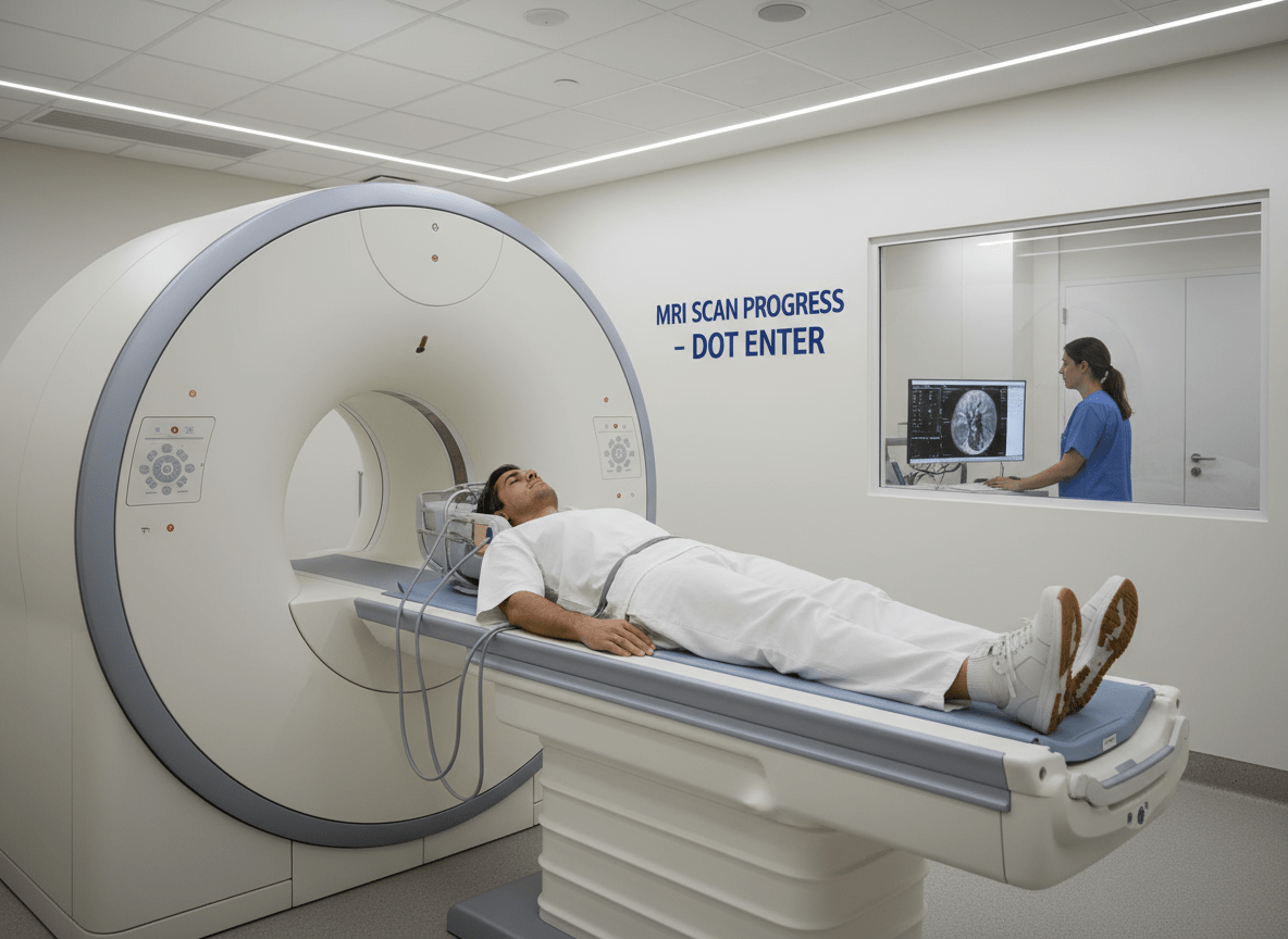
In the evolving landscape of cardiovascular imaging, cardiac magnetic resonance imaging (MRI) is emerging as a safer and equally effective alternative to single-photon emission computed tomography (SPECT) for functional diagnostics in coronary artery disease (CAD). A growing body of comparative evidence now supports what many clinicians have observed in practice: cardiac MRI can match SPECT in diagnostic accuracy without exposing patients to ionizing radiation.
Recent findings from European and international trials reinforce the equivalence of MRI and SPECT for assessing myocardial perfusion and ischemia. Studies conducted across major academic centers show that MRI provides comparable sensitivity and specificity for detecting flow-limiting coronary lesions. At the same time, MRI offers superior spatial resolution and tissue characterization, enabling clinicians to differentiate scar tissue, edema, and viable myocardium within a single session. These capabilities have expanded MRI’s clinical role beyond structural imaging into the realm of comprehensive, radiation-free functional assessment.
What You Need To Know
- Cardiac MRI matches SPECT in diagnostic accuracy for detecting ischemia and perfusion deficits in coronary artery disease, offering comparable sensitivity and specificity.
- MRI eliminates radiation exposure, providing a safer option for patients—particularly younger individuals and those requiring repeated imaging for chronic cardiac conditions.
- Technological advances such as AI-assisted quantification, faster pulse sequences, and dedicated cardiac coils are improving MRI efficiency, accessibility, and reproducibility.
- Clinical adoption is growing as updated cardiology guidelines recognize MRI’s diagnostic and prognostic value, positioning it as a key tool in value-based, noninvasive cardiac care.
Unlike SPECT, which relies on radioactive tracers, cardiac MRI uses magnetic fields and radiofrequency pulses to generate detailed images of cardiac tissue. This radiation-free profile makes MRI particularly advantageous for younger patients and those requiring repeated follow-up imaging. In populations with chronic conditions such as stable angina or ischemic cardiomyopathy, where serial scans may be necessary to track disease progression or therapy response, minimizing radiation exposure is a meaningful step toward safer long-term care.
From a diagnostic workflow perspective, the evolution of cardiac MRI has been driven by advances in scanner technology, faster pulse sequences, and automated analysis software. Techniques such as quantitative perfusion mapping and stress perfusion MRI have streamlined acquisition times and improved reproducibility. As a result, MRI is now capable of delivering both anatomical and functional information within a 30–45 minute protocol an efficiency milestone that brings it closer to routine clinical use.
Clinicians also highlight the expanding evidence base supporting MRI’s prognostic power. Data from multicenter trials and registry analyses indicate that abnormal stress MRI findings are strongly predictive of adverse cardiovascular events, comparable to or exceeding those derived from nuclear imaging. This prognostic correlation strengthens the case for MRI not just as an alternative to SPECT, but as a central tool in personalized cardiac risk stratification.
However, widespread adoption of cardiac MRI still faces structural and operational hurdles. Scanner availability, examination cost, and the need for specialized radiological expertise continue to limit access, particularly outside tertiary centers. In contrast, SPECT remains more broadly deployed in community settings, backed by decades of operational familiarity and reimbursement infrastructure. Bridging this gap will require both technology dissemination and payment model adaptation to recognize MRI’s long-term safety and cost advantages.
MedTech innovators are already responding. Vendors are introducing AI-assisted post-processing tools that automate perfusion quantification and reduce interpretation variability. The latest generation of 1.5T and 3T MRI systems integrates cardiac-specific coils and faster reconstruction algorithms, significantly improving image quality at reduced scan times. These advances, coupled with the absence of radiotracer supply chains, could help MRI gain traction in value-based cardiac imaging pathways.
Regulatory and professional bodies are also paying closer attention. Updated European and U.S. cardiology guidelines increasingly cite MRI as a first-line or complementary modality in the assessment of ischemic heart disease, especially when radiation exposure is a concern. As evidence continues to accumulate, MRI may gradually move from a niche diagnostic tool to a cornerstone of noninvasive cardiac evaluation.
While SPECT remains a workhorse of nuclear cardiology, the balance of innovation is tilting. Cardiac MRI once viewed as a specialized research modality is steadily gaining ground as a mainstream, patient-friendly alternative. The next phase will depend on continued investment in access, training, and evidence generation to translate technical capability into routine clinical impact.
For media inquiries or to share perspectives on access and evidence policy, email editor@synopulse.com.