
Tau Binds a Complex I Subunit and Reverses Electron Flow, With No Tangles Required
The case against tau has rested on two things for three decades: it aggregates into neurofibrillary tangles, and in health it stabilises microtubules. A Stanford Medicine team reports a third mechanism that requires neither, running instead through the mitochondria.
Mechanistic study across four systems: fruit flies, mice, post-mortem human brain tissue and hiPSC-derived neurons carrying pathogenic tau mutations, plus a gene-duplication model of accelerated Alzheimer’s. Senior author Bingwei Lu, with Wen Li and Suman Rimal sharing lead authorship. Published in Neuron, 6 August 2026.
- Tau molecules that acquire particular phosphorylation events cross into mitochondria. Of the roughly 80 phosphorylation sites a single tau molecule can carry, only some grant entry.
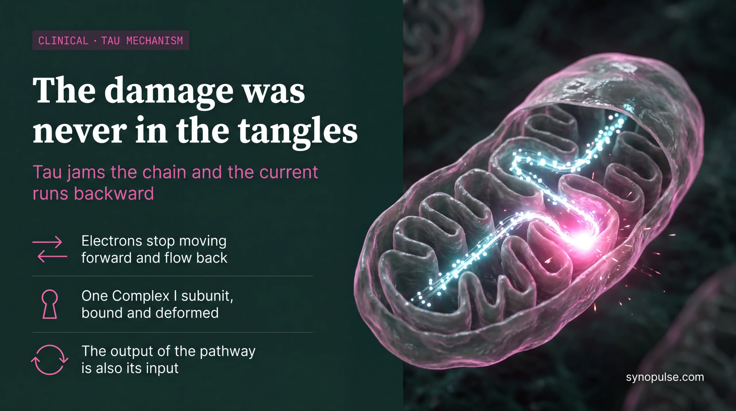
- Once inside, tau binds NDUFS3, a core subunit of Complex I, and distorts its shape. Electrons stop moving forward along the transport chain and begin flowing backward, a state known as reverse electron transport.
- The reversal generates reactive oxygen species that damage proteins, drive inflammation and phosphorylate further tau, feeding the cycle again. The pathway operated independent of both tangle formation and microtubule instability.
- Animals engineered to produce little or no tau were spared the nervous-system damage and behavioural deficits that genetically identical tau-producing animals suffered under stress. Reverse electron transport itself was first described in the 1960s and has no established constructive physiological role, with the authors reporting very little of it in healthy cells and activation under stress.
- CPT, an experimental compound, blocked tau binding to NDUFS3 without impairing normal forward flow. In tauopathy mice with established cognitive deficits, an extended regimen suppressed reverse electron transport in brain mitochondria, improved behavioural performance, prevented neuroinflammation and preserved cortical thickness and total brain volume.
Tau-PET images aggregated tau. Plasma p-tau assays have become the screening layer because they track that same pathology. Both are measuring deposits.
If the damaging species is a soluble phosphorylated tau operating inside mitochondria, tangle burden is a correlate of the disease process rather than the process itself. An asset could cut measurable aggregate and leave the pathway running. An asset could shut the pathway down and barely move tangle burden. Both outcomes read as failure or noise against endpoints built on the aggregation model.
The question worth putting to every tau programme now in development is not whether it lowers tau or clears tangles, but whether anyone has measured what it does to mitochondrial electron flow. Most will not know.